NOVEMBER 2016
I am going to undergo a Visian ICl procedure and decided to write a post about my decision. I have myopia of -2 in my right eye and -1.75 in my left eye. For those that are considering the procedure, here are my thoughts.
What is Visian ICL
The Visian ICL is made from Collamer, a biocompatible material created and used exclusively by STAAR Surgical. This lens is able to treat a large range of refractive error varying by region.Why have I chosen Visian ICL?
In theory Visian ICL have several advantages over Lasik.- They are removable
- They are additive vs permanent
- Perhaps better outcome predictability
- If something goes wrong they can be removed
As with any surgery, there are risk associated with Visian ICL over lasik. As usual I am going to scrutinize every step of the procedure.
Risks associated with Visian ICL
My main concern is that the long term effects of these lenses remains unquantified. The side effects are cataract formation, endothelial cell loss, glaucoma, increased pressure inside the eye, inflammation and other less extreme side effects such as hallos and starbursts.#1 risk with Visian ICL — cataract
My biggest worry is the early formation of cataract. When researching about the cataract formation, I went to two different sources the website of STAAR surgical (here is a brochure about them) and lasikcomplications.com. Basically cataract formation goes from 13% after 5 years to 0 on their promoted material.Encouraging is that the marketing material states this.
“Cataract. In the studies that specifically reported the inci- dence of adverse events after EVO Visian ICL implantation, which included 1,291 eyes followed for up to 5 years, there was a zero incidence of ICL-related lens opacity, visually significant cataract, pupillary block, and pigment dispersion glaucoma. In one study that extended beyond 5 years, Brar et al38 showed that, in a population of 957 eyes (V4, 615; EVO, 342), explanation due to an ICL-related cataract was only required in four cases. All of these were with the older model of the Visian ICL. Given the results of the literature review, described in detail above, it is clear that the addition of the KS-Aquaport to the Visian ICL does not alter the effectiveness of the lens. In fact, it provides equivalent quality of vision and safety compared to the FDA-approved model, without the require- ment for preoperative iridotomies.” — http://www.a-a-m.de/fileadmin/templates/augenklinik/pdf/CRST-Supplement-Sep_2016.pdf
The Staar website says;
“that the incidence of Catarakt formation is Complications After 1st Week: increase in astigmatism (0.4%), loss of best corrected vision (1.9%), clouding of the crystalline lens (cataract,1.4%), loss of cells from the back surface of the cornea responsible for the cornea remaining clear (endothelial cell loss, 8.9% at 3 years), increase in eye pressure (0.4%), iris prolapse (0.2%), cloudy areas on the crystalline lens that may or may not cause patient symptoms (lens opacities, 2.7%), macular hemorrhage (0.2%), retinal detachment (0.6%), secondary Visian ICL related surgeries (replacements, repositionings, removals, removals with cataract extraction, 3.1%), subretinal hemorrhage (0.2%), too much or too little nearsightedness correction (2.0% off by more than 2 D), and additional YAG iridotomy (3.2%).” — http://staar.com/file/10-0001-41-Rev-C-1.pdf
The prospect further goes and states, “The long-term rate of cataract formation (decrease clarity of your natural crystalline lens) secondary to implantation / removal and/or replacement of the Visian ICL are unknown.”
Of course when I visited the website — http://lasikcomplications.com/implantable-lens-risks-and-complications.htm they stated
“Moya et al. Implantable Collamer Lens for Myopia: Assessment 12 Years After Implantation. J Refract Surg. 2015 Aug;31(8):548–56.
9% of patients lost more than two lines of vision on the eye chart with glasses. In other words, if the patient could be corrected to 20/20 before ICL surgery, the patient could only see 20/30 or worse with glasses 12 years
13.88% of patients developed clinically relevant cataracts [and many more developed milder cataracts].
Endothelial cell density decreased by 19.75%. Endothelial cells are the cells on the back of the cornea that keep the cornea clear. They do not regenerate. If too many cells are lost, the patient may face corneal transplant. Link to abstract
Of course this made me very worried.
Lets get rational and get to meta-studies.
Normal population
The association of increasing cataract with age is expected, with very large relative risk found with advancing age in multiple epidemiological studies.45–48 The overall frequency of visually significant cataract in the worse eye of patients without a history of intraocular surgery aged 43–54 has been reported as 1.6% (2.6% in women and 0.4% in men).49
Visian ICL population
Table 3 summarizes information on the incidence of lens opacities and cataract surgery from long-term studies of ICL implantation. In the context of these ten publications, which present data on 5,477 eyes followed for up to 10 years, the relatively higher incidences of opacities and cataract surgery in the reports by Schmidinger17 and Guber56 (n=159) appear to represent outliers. Excluding these reports, the incidence of ASC opacities ranges from 1.1% to 5.9%, and the incidence of ASC cataracts requiring surgery ranges from 0% to 1.8%. Higher myopia in Schmidinger et al,17 and a relatively older population and higher myopia in Guber et al,56 may help explain their higher rates of reported lens opacities and cataracts.What helped me to decide that this is no longer a risk is the central hole, develped in Japan and tested on the pigs. https://www.ncbi.nlm.nih.gov/pmc/articles/PMC2292472/ they implanted pigs with the Visian ICL with a hole inside and non of them developed cataracts and with the previous version 4 all of them developed cataracts at mark 3 months.
Makes me think that the hole helps the circulation of aquaeous humor inside the eye and helps to clean the cristalline lense.
The MTF of the perforated ICL (hole diameter, 1.0 mm) in the center of the optic resembled that of the unperforated ICL. In all cases with non-perforated ICLs, subcapsular turbidity developed, but no staining caused by EB was observed in the anterior capsule. On the other hand, the anterior capsules of the eyes fitted with ICLs with a 1.0-mm hole were stained, but exhibited no turbidity.
 | |
|
Fujisawa et al. have reported that ICLs with a 3.0 mm hole in the center of the optic improved the aqueous humor circulation, and that there was no turbidity in any instance beneath the anterior capsule, but they did not examine the optical character of the lens [2].
The mean percentage of endothelial cell loss was 3.3% 10 years after surgery in the present study, which was low in comparison with that in other previous studies. Jiménez-Alfaro et al. reported that the percentage of endothelial cell loss was 6.6% 2 years after surgery.3 The U.S. FDA trial demonstrated that it was 8.4% to 9.7% 3 years postoperatively.9 Lackner et al. stated that the endothelial cell density was slightly decreased in eyes with a clear lens, but that the decrease was more pronounced in those that developed pacification. — http://mlib.kitasato-u.ac.jp/homepage/ktms/kaishi/pdf/KMJ42-2/KMJ42-2p111-119.pdf
The normal rate of endothelial cell loss is. he monolayer of cells called the corneal endothelium that lines the posterior corneal surface is derived from the neural crest during embryologic development.1 Human endothelial cell density is approximately 6000 cells/mm2 during the first month of life,2 but decreases to about 3500 cells/mm2 by age 5 years.3 Growth of the cornea accounts for some of this decrease in density, but a decrease in the number of cells also occurs.2 There is no evidence that human endothelial cells divide under normal circumstances, although they can be induced to divide in cultured corneas.4,5 They are arrested in the G1 phase of the cell cycle.6 The reason for the lack of progression into the cell cycle is still unknown, but it may involve cellular contact inhibition.7 Central endothelial cell density decreases throughout life at an average rate of about 0.6%/year8 so that the mean cell density was found to decrease from 3400 cells/mm2 at age 15 to 2300 cells/mm2 at age 85 years.9 Racial or geographic differences appear to exist; higher cell densities were found in Japanese than in American subjects.10
When you have a low count the retinal will begin clouding.
If this happens i would go for the removal hope that the eye is still fine with everything.
#2 biggest problem — endothelial cell loss
second worry is endothelial cell loss. When you loose enough of these cell, you would need a corneal transplant. And I am too young to get a corneal transplant. I think it might be cause of the barrier between the natural eye and the other part of the eye. The treatment is to get a corneal transplant however the success rate is rather low, only 30%.The mean percentage of endothelial cell loss was 3.3% 10 years after surgery in the present study, which was low in comparison with that in other previous studies. Jiménez-Alfaro et al. reported that the percentage of endothelial cell loss was 6.6% 2 years after surgery.3 The U.S. FDA trial demonstrated that it was 8.4% to 9.7% 3 years postoperatively.9 Lackner et al. stated that the endothelial cell density was slightly decreased in eyes with a clear lens, but that the decrease was more pronounced in those that developed pacification. — http://mlib.kitasato-u.ac.jp/homepage/ktms/kaishi/pdf/KMJ42-2/KMJ42-2p111-119.pdf
The normal rate of endothelial cell loss is. he monolayer of cells called the corneal endothelium that lines the posterior corneal surface is derived from the neural crest during embryologic development.1 Human endothelial cell density is approximately 6000 cells/mm2 during the first month of life,2 but decreases to about 3500 cells/mm2 by age 5 years.3 Growth of the cornea accounts for some of this decrease in density, but a decrease in the number of cells also occurs.2 There is no evidence that human endothelial cells divide under normal circumstances, although they can be induced to divide in cultured corneas.4,5 They are arrested in the G1 phase of the cell cycle.6 The reason for the lack of progression into the cell cycle is still unknown, but it may involve cellular contact inhibition.7 Central endothelial cell density decreases throughout life at an average rate of about 0.6%/year8 so that the mean cell density was found to decrease from 3400 cells/mm2 at age 15 to 2300 cells/mm2 at age 85 years.9 Racial or geographic differences appear to exist; higher cell densities were found in Japanese than in American subjects.10
When you have a low count the retinal will begin clouding.
#3 biggest problem — increased inter ocular pressure leading to glaucoma
If the lenses is not properly sized, size is key it might create pressure towards the iris and cause the increased pressure leading to glaucoma impairing the vision.If this happens i would go for the removal hope that the eye is still fine with everything.
#4 retinal detachment
Retinal detachment might occur as a result of trauma to the eye end increased pressure. It can be seen as flashes, dark shadow in the corner of the eye and floaters. Apparently the incidence is only 1 percent but still. A very serious complications. Hmm the doctor told me that if you have dilated pupil and the surgeon looks inside he should see blood and other debree also this happens in people with predisposition that the pressure caused by viscoelastic or something can trigger the retinal tear, it is a very fine material and can happen, Usually it can be treated with laser or some freezing but anyhow it is very serious. It can happen with people who have distrophy.#5 de-pigmentation of iris
Acute depigmentation of the iris is a new clinical condition that is characterized by an acute onset of pigment dispersion in the anterior chamber, depigmentation and discoloration of the iris stroma, pigment deposition in the anterior chamber angle, and positive transilluminationSo finally i went to get the surgery.
Surgery
#1 Day with Visian ICL
Day one I’ve come to the clinic in the morning everything is going fine the surgery. Well first of all they put bunch of drops into your eyes so you don’t feel anything and then you undergo the surgery. The surgery itself is not very comfortable but it’s painless before the surgery the surgeon talks to you.I asked him how did he calculate the space between the crystalline lens, he said that the Visian ICL is creating the space between the two.
He told me that after the surgery about 40% of people will have increased intra-ocular pressure therefore they might put some drops to reduce the pressure and I might get a migraine.
So I go to have the surgery first, you start looking into a very bright light which is very uncomfortable but you get used to it and then they inject this substance-viscoelastic into your eyes which makes it totally blurry and you can actually see the contours of the knife penetrating your eye.
The most uncomfortable part is when he tries to put the Visian ICL in which feels like pulling off the eye sideways. And after the surgery he injects some sort of agent to constrict-phylocarpine the pupil. After the surgery everything seems to be hazy and my pupil is constricted and I get a headache so they administer some drugs so I wait for about 23 hours and the pain slightly goes away my intraocular pressure jumps to 22 which is a bit higher but it’s nothing unusual and so I’m released to home care
#2 Day with Visian ICL
Tonight my pupil remains constricted and I don’t see very well but when I put on the glasses I can see quite well everything seems to be dark orange. Feels like sand in the eye, it keeps tearing. In the morning I go for the checkup and the doctor tells me that they put a lot of agent inside so my pupil remains constricted. I’m quite not happy with the outcome because everything seems to be hazy, cloudy I don’t see well and I can see circles around light even the sunlight. Entire day goes like this. I am starting to read about non responsive pupil. Which can happen to some people and it is a permanent condition. I have not considered as a risk factor in the surgery.#3 Day with Visian ICL
On the third day I’m supposed to have the surgery in the second. I noticed there is a shadow in the corner of my peripheral vision of the eye that was operated on.Immediately, I start freaking out about retinal detachment, retinal tear so when I go for the check up after the surgery, I refused to undergo the second surgery until my first eye is completely fine, the doctor wasn’t happy he told me he has 16 surgeries this day and he’s only waiting for me my pupil remains constricted I cannot see very well everything reminds dark orange. They open my eye they put the agent to make the pupil big and they looked into my eye saying that the Visian ICL is sitting right in the middle and that the shadow is some sort of reflection from the lens. This happens when I look closer to my nose it’s like something is in my periphery and I also see the circles around lights so he looks into my eyes and says that everything looks to be fine but still I refuse to go for my second surgery.
Apparently the retinal detachment or a tear demonstrates in flashes. He looked into my eye with the slit lap and hope he did not see any any retinal stuff happening.
#4 Day with Visian ICL
On the fourth day my pupil dilates a little bit. I am seeing better than the other two days but I still don’t see perfectly as with the contact lenses or with my glasses, I can observe that my eye changed color and also that the iris is bulged out on the eye that was operated on I have a fear they missed calculated the size of the lens which may lead to that pigmentation of the iris or increase intra-ocular pressure leading to glaucoma. | |
|
http://www.healio.com/ophthalmology/journals/jrs/2014-3-30-3/%7B4f00e2a0-d1de-4572-8e6b-10e2fa7e59f6%7D/measurement-of-the-crystalline-lens-radius-with-artemis-very-high-frequency-ultrasound-biomicroscopy-for-implantable-collamer-lens-sizing.pdf — Measurement of the Crystalline Lens Radius With Artemis Very High Frequency Ultrasound Biomicroscopy for Implantable Collamer Lens Sizing.
#5 Day with Visian ICL
My pupil is still not ok, it is still constricted and when exposed to light does not constricts as much as the other one. I am going have to wait a bit. Also, I am feeling a bit of pressure in my head a head ache, albeit a light one. Is my inter ocular pressure building as a result of papillary block? The vision has improved, yet I still don’t see as with the other eye with glasses. The shadow on my peripheral vision is still there, extremely annoying. What could it be.- retinal tear — but would not they see it
- a play of light
- as a result of bulging i am seeing different things at the corner of my eyes, eyelashes and stuff.
- there is some sort of pressure from the visian icl at certain points that is causing the dark spot to appear.
I was doing some work with computer and checking the numbers and invoices and facts and the pressure seems to building up. I am not convinced and will not undergo another surgery next Tuesday.
Still seeing circles around lights, which is rather funny. But the dark spot on in the left corner of my eye is worrisome at least.

Vision with Visian ICL at day 5. Seeing circles that should go away once the brain filters them out, I am more worried about the dark spot. Further I go with my eye to the right side to my nose more pronounced it is forming some sort of orb.
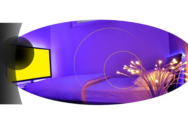

#6 Day with visian ICL
I am not happy with the outcome, not only my vision is not perfect, but i am experiencing annoying side effects. Stuff that worries me.1, shadow in the corner of my eye — could i experience the famous retinal detachment
2, bulging of the iris
3, slight headache — could the IOP building up?
4, light scratching when i look up right
5, Still not seeing sharp as with glasses
6, My pupil is still not working properly.
7, circles
The dark spot starts change color to light to transparent or flashing, it also changes if i am closer to the mirror, thus i suspect that it is the lens itself and not a retinal detachment. I will see if this persists or goes away with neuro-adaptation of the eye.
When i look towards the light my un-operated eye is perfectly clear but the operated eye contains a few floaters. Not as many as below image but one with two dots. Plus some like curtains looks like a gel that is being floated around and casts very subtle shades. Hope this condition will go away with neuroadaptation. The dark circle in the perifery might also go away with neuroadaptation. My vision is still not super as with classes but have improved since monday.
I noticed a few floaters like one and a dot similar to below, it can be a sign of cobwebs from retina or something else, the literature suggest they should go away. While my unoperated eye is cristal clear my Visian ICL eye when i look into the eye i can see the two floaters and also some sort of gellike substances that are throwing shades, seems like some sort of debree from the viscoelastic or some sort of debree from the surgery. Hope they will go away as the literature suggest they will go away.
 |
| http://www.eyereview.com/floaters_after_cataract_surgery.html |
 |
| Floaters following Visian ICL surgery |
#7 Day with Visian ICL
My distance vision has improved, i think I might have like -0,25 im my operated eye. I still see the dark circle in the corner of my eye.I noticed that my operated eye has changed color??? What is going on?
 |
| Notice the change in my eye color |
 |
right eye is with the visian icl is darker and has larger pupil in the same light condition as opposed to my non Visian ICL one.
|
Not that the bulding of the eyes could be cause the visian ICL is actually lifting the iris. Here is image I found on the Internet.
 |
you can see the the visian ICL is lifting the iris, thus chaning the color of your eye.
|
 | |
| This picture is inaccurate the Visian ICL does press against the iris….look at the change in color of my eyes and the fact that the |
My eyes used to be lighter in color now the operated eye has dark blue color instead of the light gray. You can see the my unoperated eye has the ligher color. Why is it changing?
I suspect the my Visian ICL is pressing against the iris and is creating the bulging effect thus the light is being dispersed in the different way. I am not very happy that my iris has changed color. Hope this is not an indication of the wrong sizing as it might lead to higher inter ocular pressure, glaucoma and eventually pigment dispersion.
The functionality of my iris is getting hopefully restored. I occasionally feel pressure in my head.
Also I noticed that I have problems with accommodation in the operated eye. It seems that my near distance vision is worse, I can see so sharp with my unoperated eye, but with my operated eye near distance text, or small print is doubling and i have to really focus to read something. This has not been reported in the side effects, that it might impact your near distance vision. If you are working with computers it can have negative effects.
I am still undecided on the second surgery, albeit it has improved a lot.
Also, i have to report that the night vision has worsen, i can see less light at night in my operated eye as opposed to my unoperated eye. It seems to be related to the iris dilatation, as the eye with Visian ICL is not dilating as opposed to the operated eye. It can be because my eye overreacted to the constriction, I dont know what have i been given at the end of the Visian ICL surgery and is still getting used to. In the intereted they reported 7 days for the iris to start functioning as before. Some have reported that their iris is being permanently dilated — so called http://www.djo.harvard.edu/site.php?url=/physicians/cr/2755
We describe the case of a healthy 28-year-old woman who underwent implantation of a posterior chamber, phakic, toric, implantable Collamer lens (ICL) in both eyes for correction of bilateral high myopia with astigmatism. On the first postoperative day the patient developed increased intraocular pressure (IOP) and a fixed, mid-dilated pupil in her left eye. The elevated IOP was corrected within 3 days by medical treatment. However, the pupil remained mid-dilated and nonreactive to both light and accommodative effort during 2 months of follow-up; there was no reaction to pilocarpine (0.125% or 4%) eyedrops. The patient was diagnosed with Urrets-Zavalia syndrome (UZS), which has been reported in association with ICL implantation only once previously.
Hmm, but the opposite such as in my case that the iris does not want to open up is not very common.
Also I noticed the change in my iris hole, the operated eye has a slightly oval shape in the evening, dim light settings than that of the unoperated eye. It seems that following the Visian ICL surgery my pupil when from round to oval, perhaps that is the cause of the shadow at the corner of my eye.I suspect the my Visian ICL is pressing against the iris and is creating the bulging effect thus the light is being dispersed in the different way. I am not very happy that my iris has changed color. Hope this is not an indication of the wrong sizing as it might lead to higher inter ocular pressure, glaucoma and eventually pigment dispersion.
The functionality of my iris is getting hopefully restored. I occasionally feel pressure in my head.
Also I noticed that I have problems with accommodation in the operated eye. It seems that my near distance vision is worse, I can see so sharp with my unoperated eye, but with my operated eye near distance text, or small print is doubling and i have to really focus to read something. This has not been reported in the side effects, that it might impact your near distance vision. If you are working with computers it can have negative effects.
I am still undecided on the second surgery, albeit it has improved a lot.
Also, i have to report that the night vision has worsen, i can see less light at night in my operated eye as opposed to my unoperated eye. It seems to be related to the iris dilatation, as the eye with Visian ICL is not dilating as opposed to the operated eye. It can be because my eye overreacted to the constriction, I dont know what have i been given at the end of the Visian ICL surgery and is still getting used to. In the intereted they reported 7 days for the iris to start functioning as before. Some have reported that their iris is being permanently dilated — so called http://www.djo.harvard.edu/site.php?url=/physicians/cr/2755
We describe the case of a healthy 28-year-old woman who underwent implantation of a posterior chamber, phakic, toric, implantable Collamer lens (ICL) in both eyes for correction of bilateral high myopia with astigmatism. On the first postoperative day the patient developed increased intraocular pressure (IOP) and a fixed, mid-dilated pupil in her left eye. The elevated IOP was corrected within 3 days by medical treatment. However, the pupil remained mid-dilated and nonreactive to both light and accommodative effort during 2 months of follow-up; there was no reaction to pilocarpine (0.125% or 4%) eyedrops. The patient was diagnosed with Urrets-Zavalia syndrome (UZS), which has been reported in association with ICL implantation only once previously.
Hmm, but the opposite such as in my case that the iris does not want to open up is not very common.
Here the image — you can see that my eye does not dilate in the low light settings as before. Even in the image it is clear that the eye without the Visian ICL surgery has larger pupil — even tough it is exposed to more light.
My worries at day 7 with Visian ICL
- low light settings and iris functioning
- change in the color of the eye
- accommodation and near sight vision
- pressure in my head when i try to work close distance, computer or devices.
We can see from the picture above that my pupil has changed shape from circular round shape to more oval shape. Not sure what to make of it. Searching the Internet i can come up to this conclusion.
The only response i have is the below.
Dr. Holland notes that another concern has been ovalization of the pupil caused by the pressure of the haptics pushing out into the angle. “In the AcrySof trials, out of more than 700 eyes implanted with the lens, there have been no cases of pupil ovalization,” he says. “That’s unusual with such a large cohort. It may be because the haptic loops on this lens are flexible; if tissue is pressing against the loop, the loop flattens a little into an oval shape. This makes precise sizing less of an issue than it is with posterior chamber phakics; we just use white-to-white, which is inaccurate, but we haven’t had any issues as a result.”
An oval pupil can occur, regardless of all the precautions that might have been taken at the time of surgery. Round pupils at the time of surgery and the confirmation of the correct placement of the feet by intraoperative gonioscopy may not prevent the occurrence of pupil ovalization later on. An oval pupil as such produces no symptoms. However, if the process of ovalization continues to increase optical problems, low-grade uveitis and decentration can occur. The late ovalization of the original round pupil is caused by callous formation where the feet of the implant impinges on the iris. The callous formation can contract, causing a progressive pupillary distortion.
. Regarding the change of the color in my operated guy the doctor said the ICL is pushing the iris outwards us as the iris rests on the ICL.She said that this condition is very normal.
I talked about different light sensitivity with the ICL in the unoperated eye. And also I told her about ovalization of my pupil. And also I told her about ovalisation of pupil. She did not find any inconsistencies she said that my pupil is reacting well to the light and she told me that it is perfectly round she said that the slight imperfection can be present I ask her if my ICL could be pressing against the angle as it might be too large for the eye I ask her if my ICL could be pressing against The corner of the iris. and asked her if I should do the ultrasound exam to which she replied that ultrasound is not very precise instead see she suggested I should do the exam on pentacam and asked her if I should do the ultrasound exam to which she replied that ultrasound is not very precise instead see she suggested I should do the exam on pentacam and oct, The pentacam showed that my valut is about 3,2 mm which is not bulging forward. With the oct, they have the older model we could not see the footplates of the visian icl but we could se the valut. They . reassured me that from the medical perspective everything looks fine and let me decide if I want to do the other I are I want the removal
I decided to go forward with the second eye and try to do the surgery in the second eye.
Currently my eye is in pain especially the upper part where the incision was and i Experience headache.
After the Visian ICL surgery, the eye would really burn, cut and the feeling of having sand in my eye was omnipresent. It was very unconfortable and I could not do anything in the evening. It was constantly tearing and had discharge from my nose. When I woke up it was fine, they eye was just super red.
I went to the doctor for the check up.
She said that everything looks good and had a few questions for her. My pressure was 8 and 11 in left and right eye.
1, can i play beach volley with Visian ICL
she said it will take 2 weeks
2, can i go to the gym with Visian ICL
she said I should wait for 2 weeks
3, can i go to smoke places and drink with Visian ICL
she said I can but need to hydrate my eye
4, can i rub my eye with Visian ICL
should not do that now but in two weeks i can, apparntly the cornea is strong to withstand the pressure, so the visian icl will not touch the cristalline lense. The problem might be paint ball or golf a direct impact into the eye.
5, how can we prevent cataract formation, glaucoma or iris depigmentation with Visian ICL
Well time will tell if this surgery is successful
My right eye is much better the iris is constricted but i can see reasonably well than with my left eye.
I also talked about the intraocular surgery risk associated with Visian ICL, any intraocular surgery has risk as you are tempering with a closed very intricate system, there are many things that might go wrong, you touch the crystaline lense, develop cataract, your iris can stop functioning as there might be reaction to medication, you can cause retinal tear with high pressure, you can induce glaucauma, the surgerys long term negative effects can manifest in years to come. The eye is not something that is designed to be entered. With all these negative I feel that the laser surgery is better in a way as everything happens on the cornea and not inside the eye, but it is permanent.
Apparently — ICL implantation increases corneal astigmatism by a with-the-rule shift of approximately 0.50 D. Although this is a small change, it may not be negligible; refractive surgery such as LASIK or toric phakic IOL implantation should aim to fully correct spherical and cylindrical error. — http://crstodayeurope.com/articles/2010-nov/feature-story-surgically-induced-astigmatism-after-visian-icl-implantation/
This is my vision at night with an iphone.
Dr. Holland notes that another concern has been ovalization of the pupil caused by the pressure of the haptics pushing out into the angle. “In the AcrySof trials, out of more than 700 eyes implanted with the lens, there have been no cases of pupil ovalization,” he says. “That’s unusual with such a large cohort. It may be because the haptic loops on this lens are flexible; if tissue is pressing against the loop, the loop flattens a little into an oval shape. This makes precise sizing less of an issue than it is with posterior chamber phakics; we just use white-to-white, which is inaccurate, but we haven’t had any issues as a result.”
An oval pupil can occur, regardless of all the precautions that might have been taken at the time of surgery. Round pupils at the time of surgery and the confirmation of the correct placement of the feet by intraoperative gonioscopy may not prevent the occurrence of pupil ovalization later on. An oval pupil as such produces no symptoms. However, if the process of ovalization continues to increase optical problems, low-grade uveitis and decentration can occur. The late ovalization of the original round pupil is caused by callous formation where the feet of the implant impinges on the iris. The callous formation can contract, causing a progressive pupillary distortion.
#8 and 9 with Visian ICL
I’ve just had the second surgery in my right eye the duration of the surgery was about 15 minutes it wasn’t painful but very uncomfortable as you have to look into a very bright light and you feel pulling and pushing towards your I also division gets really cloudy and you can see the blades are the contours of the blades and you can also see the vehicle elastic coming into your eye after about 20 minutes I developed slight headache it’s not too much on my I pressure job to 17 I was waiting at the hospital for about two hours was given some drops to lower my pressure and pain killers and I was released to home.. Regarding the change of the color in my operated guy the doctor said the ICL is pushing the iris outwards us as the iris rests on the ICL.She said that this condition is very normal.
I talked about different light sensitivity with the ICL in the unoperated eye. And also I told her about ovalization of my pupil. And also I told her about ovalisation of pupil. She did not find any inconsistencies she said that my pupil is reacting well to the light and she told me that it is perfectly round she said that the slight imperfection can be present I ask her if my ICL could be pressing against the angle as it might be too large for the eye I ask her if my ICL could be pressing against The corner of the iris. and asked her if I should do the ultrasound exam to which she replied that ultrasound is not very precise instead see she suggested I should do the exam on pentacam and asked her if I should do the ultrasound exam to which she replied that ultrasound is not very precise instead see she suggested I should do the exam on pentacam and oct, The pentacam showed that my valut is about 3,2 mm which is not bulging forward. With the oct, they have the older model we could not see the footplates of the visian icl but we could se the valut. They . reassured me that from the medical perspective everything looks fine and let me decide if I want to do the other I are I want the removal
I decided to go forward with the second eye and try to do the surgery in the second eye.
Currently my eye is in pain especially the upper part where the incision was and i Experience headache.
After the Visian ICL surgery, the eye would really burn, cut and the feeling of having sand in my eye was omnipresent. It was very unconfortable and I could not do anything in the evening. It was constantly tearing and had discharge from my nose. When I woke up it was fine, they eye was just super red.
I went to the doctor for the check up.
She said that everything looks good and had a few questions for her. My pressure was 8 and 11 in left and right eye.
1, can i play beach volley with Visian ICL
she said it will take 2 weeks
2, can i go to the gym with Visian ICL
she said I should wait for 2 weeks
3, can i go to smoke places and drink with Visian ICL
she said I can but need to hydrate my eye
4, can i rub my eye with Visian ICL
should not do that now but in two weeks i can, apparntly the cornea is strong to withstand the pressure, so the visian icl will not touch the cristalline lense. The problem might be paint ball or golf a direct impact into the eye.
5, how can we prevent cataract formation, glaucoma or iris depigmentation with Visian ICL
- regular visits with dilatation of the pupil will determine if i can get cataract but the OCT scan showed that the ICL is not touching the lens
- glaucoma is not an issue as the pressure is low but periodic check up should be done on yearly basis
- iris depigmentation — she looked a bit surprised by this question, they have not had a single case, but it would mean that the icl is touching the iris, she said that there is a small space between the Visian ICL and iris, some fluid is inside. And the OCT showed that the iris is not touching the Visian ICL.
Well time will tell if this surgery is successful
My right eye is much better the iris is constricted but i can see reasonably well than with my left eye.
I also talked about the intraocular surgery risk associated with Visian ICL, any intraocular surgery has risk as you are tempering with a closed very intricate system, there are many things that might go wrong, you touch the crystaline lense, develop cataract, your iris can stop functioning as there might be reaction to medication, you can cause retinal tear with high pressure, you can induce glaucauma, the surgerys long term negative effects can manifest in years to come. The eye is not something that is designed to be entered. With all these negative I feel that the laser surgery is better in a way as everything happens on the cornea and not inside the eye, but it is permanent.
#10 day with Visian ICL
Pretty uneventful my right eye is getting in shape and already can see much better the pupil is circular and everything looks fine, I am just waiting until the iris starts functioning again, as it takes a few days for the pupil to dilate. However, my left eye, during night vision it is worse has I think developed a mild form of astigmatism. I am not a doctor but the objects are a bit blurred, with my right eye everything is crystal clear no distortions. Could that be because of the ovalization of pupil?Apparently — ICL implantation increases corneal astigmatism by a with-the-rule shift of approximately 0.50 D. Although this is a small change, it may not be negligible; refractive surgery such as LASIK or toric phakic IOL implantation should aim to fully correct spherical and cylindrical error. — http://crstodayeurope.com/articles/2010-nov/feature-story-surgically-induced-astigmatism-after-visian-icl-implantation/
This is my vision at night with an iphone.
 | |
|
Hope this clears up but I don’t know.
 |
Right eye and left near vision, it flactuates but the worse I see is as displayed in the left picture. The lines are blurred, but I can still see the main text but smaller it is the more difficult is it to read. Could I have developed a mild form of astigmatism following the Visian ICL implantation and surgery?
|
Also, I think I am experiencing a a head ache as the eye is trying to focus but fails. Here is where the headache occurs following the implantation of Visian ICL lense.
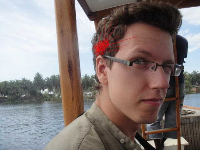 |
Red color is where the pain concentrates following the Visian ICL EVO implant in the left eye, the right eye does not experience any pain. The pain might be due to inability to foucs.
|
#11–14 Day with Visian ICL
My right eye is perfect hardly any shadow in the perifery, super focused, clear and crisp. I do get the circles. the reflections from the aquaport or the centralflow visian ICL stuff.My left eye still experiences shadow vision, or double vision and right peripheral shadow, I am also aware of the reflection in the lens in the periphery, the double shadow vision at all distances is so annoying I am getting headaches every day only in my left side. Why is this happening?
it could be because of the improper size of the ICL or tilting, or it could be that the visian icl is too big which explains why my pupil is a bit larger than in the right eye.
Could be the way how my pupil is shaped now, which as a bit of oval shape and it could be distorting the light entering the retina…
it could be because the visian icl has increased or shifted the lighfocus
Here is the night vision — with Visian ICL you can see glares and significant halos.
 |
The halos I get in my left eye from Visian ICL, the circles are normal and they are reflection of the aquaport. Its kind of artistic.
|
I contacted the https://www.arcscan.com company that produces high frequency ultrasound scanners for fitting to get a second oppinoin on my left eye visian ICL implant. I am sure that something is not quite right, although everyone seems to think it is. I just cant stand the shadow vision, its difficult to work in such condition with headaches all the time. As most surgeons use the biometric suculus to suculs exam and do not really rely on the high frequency ultrasond device as it is super expensive, I am sure they are not quite able to get the measurement right, which is a problem, for Staar Surgical. I am also considering the removal of the Visian ICL as these symptoms are quite debilitating.
Also when you place something in between your eye the peripheral shadow on the periphery grows larger with Visian ICL.
#Two weeks with Visian ICL
Right eye perfectLeft eye is the same
Main issues with the left eye:
- blurry edges, double vision
- peripheral shadow
- cobwebs, much more than in the right eye
- I feel that the left eye is more bulged
- headache and migraines because of Visian ICL in my left eye
 |
Left eye and right eye following the Visian ICL implant
|
#1 month with Visian ICL 29.1.17
It has been one month since the surgery. So I want to put the update. Well, I am getting used to the vision, the vision is generally good. The blurriness subsided a bit but I do see horizontal ghosts in my left eye, as I have been diagnosed with astigmatism in my left eye. My right eye is dominant therefore it should balance things out a bit and I should see sharp.
Hallos are still present and are the circles but I am getting used to that. I don’t think they will disappear. My doctor said that everything is OK. We shall see about that. The shadow in my left eye has subsided as well. I thing i could have seen part of my eye as your eye will change shape a bit following the surgery.
An example of my shadow vision.
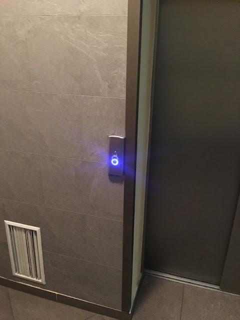 |
| Ghosting vision with Visian ICL |
When I look up close on something there are two shadows in my peripheral vision.
Also, with I close my eyes and push my eyes hard to the edge I do see dark purple shapes as I think the lens is pressing against something in the eye.
I see floaters, never seen them before, my theory is the following. Following the increased IOP during the surgery some elements are released into the vitreous gel.
Copied this from — https://www.facebook.com/groups/LasikComplicationsFaceBookGroup/search/?query=floaters
The images below are of the eye of a 25 year old patient who underwent LASIK surgery 3 years ago. These images were taken with a technology known as “OCT”( “optical coherence tomography”) and show a cross section of the retina very close to the macula. The space above the retina is where the vitreous is located which makes up the bulk of the interior of the eye. One image is in black and white and the other in color to better demonstrate the layers of the retina. About a year after undergoing LASIK, this patient noticed large amounts of floaters. He explained his vision as if looking at thousands of tiny black spots against light colored backgrounds mainly indoors. He visited a number of ophthalmologists who were unable to see or visualize anything out of the ordinary. With conventional diagnostic instruments such as a slit lamp, fundus camera or ophthalmoscope, I too was not able to see what was causing this patient to see these very tiny floaters until unitizing OCT technology. The floatersinside this patient’s eye were just too small for the human eye to detect with the instruments described above. Look carefully and note the thousands of very tiny black particles just above the retina floating within the vitreous gel that makes up the bulk of the eye. These are so close to the retina that they blend in with the retinal tissues (making it almost impossible for doctors to visualize) and cast shadows onto the retina. These tiny floaters were created by the LASIK surgery. During the LASIK surgery, the intraocular pressure is increased dramatically within a very short amount of time and then lowered in an equally dramatic manner. This rapid increase and decrease in the intraocular pressure “shakes up” the interior tissues creating this condition which may occur immediately after the surgery or years later. At the present time the only way to do away with these floatersis to perform a vitrectomy which involves removal of the vitreous ( essentially most of the contents within the eye) and replacing it with saline solution. A vitrectomy is a very risky surgery and is only done in extreme situations.
Now I am started seeing something in my left eye that looks like a eye lash but it is still there. I wonder what is happening.
Also a problem I see that there is a foreign element in the body and when you rub with your hand against your eye the crystalline lens can come in contact with the Visian ICL. Thus, you try not to do it but when I sleep sometimes I wake up with my eyes that hurt, of course i have been pressing my eye into the pillow. I hope that this contact will not set the stage for cataracts formation.
15.2.17 I stopped counting days since my Visian ICL surgery, so I would just put in the date.
I have to report a very troublesome issue. I got home from a party at 3 am in the morning my hallos got really bad in my left eye and in my right eye were quite annoying. When I got home into the room I looked at my eyes and my pupils were just huge. Plus they had uneven shape my left one, read at the begging is slightly oval, well it was super oval like a Cats eye and did not react to the light. They were just plain huge not constricting when in bright room. I do not know the cause, before I could snap a picture they started reacting again and constricted. But the fact is that they dilated beyond the Visian ICL lens and thus were causing the glare, fine the hallos are ok but the fact that they were non reactive for a while is scary. My pupil looked totally dilated with the upper edge almost completely gone oval eye as a cat and non nonresponsive.
I dont know the cause, the above is the demonstration. But I am really scared. At night when I go out and there is dim light my pupils dilate beyond the visual range of the Visian ICL, I am sure it is the result of the surgery cause they are really big. Bigger than before.
3 months following ICL Surgery — 21.2.17
Well, about two week ago I started to notice some side effects.- starbursts
- halos
- hazy vision in dim light
- larger pupil size
- left head head age as a result of astigmatism induced by the surgery
- astigmatism in the left eye following the surgery
- floaters
- random visual effects, like black spot top right in my right eye, like when i close my eyes and move them all the way to the side i see different sort of blackness in the corner meaning that the VIsian ICL is pressing against something in the eye to cause this effect.
Here in pictures:
 |
| Check how big they are, no doubt the pupil dilates beyond the visual lense. Plus they have irregular shape as a result of the iris being pushed outwards by the Visian ICL lense. |
I am not bothered by the visual effect so much as I am worried about some potential long term side effects.
I am more worried about the recent dim light or artificial light interior haziness.
When I am in dim light or artificially lighten interior, i experience hazy vision, this has not been the case about 2 weeks ago. It has started recently. I wonder what is the cause.
I will monitor the situation. It is true that the new model with aqua-port or central-flow has not been reported to cause cataracts.
- I might be developing cataract
- Neuro Adaptation, no longer do I see well defined circles, they might be more blurred and that can cause the haziness.
- it can be dry eyes and new eye drops I am using now.
- corneal endema
- loss of endothelial cells
I will monitor the situation. It is true that the new model with aqua-port or central-flow has not been reported to cause cataracts.
11.3.17 Following visian ICL surgery
I feel the floaters in my left eye are getting worse. I suppose it’s got to do with this.
The images seen below are computer enhanced cross sectional views of the right and left retinas of a 23 year old post LASIK patient. The thick green-yellow “sponge-like” structures are the layers of the retina. The semi-clear space above the retina is the vitreous which makes up the bulk of the interior of the eye. The young man had LASIK surgery done when he was 18 years old. One of his main complaints is floaters. When he looks at his computer or in a brightly lit environment he is acutely aware of them. Look carefully at the numerous dark pinpoint spots above the retina.
These are the floaters that are casting a shadow on his retinas. He is seeing the shadows and not the black spots. The shadows that he is seeing are much larger than the actual pin point dots. He was referred to a retina specialist for a consultation. A diagnosis of “Vitreous Syneresis” was made. The vitreous has a gel like consistency.
Vitreous Syneresis is characterized by a degeneration of the vitreous with a loss of gel consistency. The retinal specialist who saw this patient and I both feel that this condition is directly attributable to the LASIK surgery. We fit this patient with scleral lenses in order to enhance this young man’s vision and to treat his post-LASIK dry eyes. It is possible that at some future date, a vitrectomy (removal of the vitreous and replacement with saline solution) will need to be done. — https://www.facebook.com/groups/LasikComplicationsFaceBookGroup/
Well
26.3.17 following Visian ICL surgery
I feel that my cat eye the oval eye is getting worse, I had pretty bad problem yesterday.
I hat to flash a light inside my eye for the pupil to get to normal shape sever times a night, very stressed out about it.
I have also reevaluate my theory about the floaters. Maybe they are just the reflection from the visian ICL lens. It can be because I only see them from when on the back drop of the light reflections from the icl, like rainbows and other stuff. Maybe they would go away when removed.
I have been to the doctor. Key takeaways.
- out of 600 patients I am the only one along with some guy that is of mixed ethnicity
- this happened recently they suspect it has to do with the new design EVO V5 that is supposed to be better. No incidences of V4c
- I should contact Staar surgical for this.
- Doc. did pentacam and said that the lens is sitting nicely in the eye. She believes that some water is accumulated there and causes the upper sciliary muscle not to work properly.
- got some phylocarpine drops
- they will monitor the situation.
- apparently the condition is not dangerous. However, aesthetically it is not very pleasing and my visual effects are quite disturbing. And who knows if in the long term this cannot cause some problems.
1 year after the Visian ICL Surgery some pictures
One year after the surgery things are pretty much unchanged. I see glares, starbursts, my left eye iris still gets stuck by the edge of the lense — I suppose that is what is happening. To summarize.- when I am working a lot on the computer my pupil dilates so much in the evening i get the glares immediately when in dim light.
- when I am in the gym and do weight lifting I get the glares during the daytime as well
- when I wake up most of the time my left eye is blurry
- I was changing a tire in the evening, when I bent my head at night I get glares and my iris gets stuck leading to oval pupil as described above.
- When I talk to girls I am attracted to I get very bad glares and starburst suppose my pupil dilates from the stress and it dilates more than the visual field of the Visian ICL. My eye gets ovalized. I have to look into a light source to correct it.
- When there is reflection from the sun i see lots of circles, rainbows, stuff and on the backdrop of these white reflections I see countless floaters dots.
- At night i see massive circles when I am close to lamps.
- When I work a lot with a computer, I get blurry vision in my left eye and also headache, I suppose the lens in my left eye is at an angle causing the double vision or shadows and that is causing strain on the eye.
- GOOD news is that I dont see the peripheral shadow anymore. That have been the eyelashes and my brain is filtering them out. Circles and visual stuff not so much.
- Night life and clubbing suck as with the progress of the night there is exponential increase in glares and reflections. At 4 AM the situation becomes really bothersome.
My doc wrote Visian ICL company about my ovalized pupil and they have not responded yet. They have sent me a similar customer form but that form for the customer should win the “UX friendly customer form of the year” they pretty much do not leave any space for the customer to express problems, bunch of checkbox ex…like has there been an injury or something like that but they do not leave space for qualitative problems.
 |
not like this but dots like that and specks
|
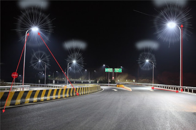 |
I see starbursts around night lights. The glare above the lamps is caused by my ovalized pupil. These glares are a lot bigger than on the picture. Night vision with Visian ICL EVO+
|
Night driving sucks
 |
Direct sun reflection with Visian ICL. Central hole creates circles and different light effects on the backdrop of which the floaters could be seen.
|
When there is reflection from the sun I see circles with some rainbow depending on the angle. On the backdrop of this reflected light there are some floaters, dots etc.
 |
Vision in the gym with Visian ICL
|
Here is the example of glares I get in the gym…usually above any object but mostly above the shiny objects. Not sure if that is to do with increased pressure. Also when i bend my head i get bad glares from Visian ICL.
 |
Vision in the gym with Visian ICL
|
Again left eye with slight shadow vision. Also when I wake up and wide open my eyes i see a dark spot in the bottom left of my vision in my left eye. In my right eye I do see it as well but not as strong….that is when I really bulge out my eye…maybe the Visian ICL is pressing against some tissue. Hope it is not the crystalline lens because it can cause cataract.
This is a great resource, the sizing is crucial in implanting IOL many clinics just dont have technology to do the sizing properly and hence the results are as they are and many problems ensue.
 |
Nightlife with Visian ICL, every source of high intensity light produces circles, in this case they are all blue as the light is blue. This is how you are going to experience clubbing. The glares are getting worse with the tiredness of eyes. If you are working all day on the computer the onset in the evening of glares is faster than when the eye is rested.
|
23.2.2018 Comments
Here is the great resource - https://www.sedesoi.com/leggi_premi_archivio.php?area=ricerca&pagina=lecture&id=1181&fbclid=IwAR0yTpdT_PtKoWFfbLgXRspFUgUuASSsy05ghoMZbUTrZ0Sa4PLCGuqjpdQ
The doctor is however consultant for Staar Surgical...
Sizing is still a weak point of the ICL, he said. A wrong vault can cause cataract, and with current methods of sizing, the spread of results is high.
“Vault is the most important thing in ICL. The lens has big tolerance in sizing, but you need to know how the vault works. What appears to be an adequate vault may no longer be so when the pupil constricts. People living in bright light conditions may have for a long time hypoxia in the capsule and develop cataract, even with the central hole,” Zaldivar said.
-
5.1.2019
https://www.facebook.com/VisianICL/ the facebook page turned off their reviews...too negative????


Hello ! Hope that you will feel ok and see perfect !
ReplyDeletePlease tell us, what exact model of the VISIAN you have in your eyes? Please post updates. Tell us if you feel dye eye.
I do not understand why u did this for only -2 ? I have-12…..
If i have -2 I was the happiest person in this world !
Hi thanks for you message, I have the latest model it is V5c or they call it EVO.
DeleteThere are several reasons why i did it. It is reversible, the rational for that is that I would have it removed in 10 years and will not to need the reading glasses when I wont do as much sports. It seemed less invasive as it is an additive not permanent solution unlike Lasik.
Maybe I fell for the marketing ploy, I dont know. I do have dry eye in my left eye but the right one is good.
Hello !
ReplyDeleteI did not get this pharese : “i n 10 years and will not to need the reading glasses” How is that ? The accomodation and cataract depends on your eye ( genetics ). Maybe in 10 years or 15 you will need IOL lense exachange with an artificial crystalin.
How do you feel/see ?
Do not forget to post :)
Well they told me “the doctors” that i will not need reading glasses with mild myopia, unlike people that have 20/20 vision with Lasik, who would need reading glasses with they reach certain age. I wanted to have this option open for me.
ReplyDeleteI would remove them regardless of getting cataract. I have not read about getting cataracts with the new aqua-port version but perhaps the studies are quite recent.
As for the vision halos started to appear 3 months post op and i see starburst as my eye started to dilate quite a bit. Also, it seems that my vision is getting a bit hazy in dim light.
Hello Peter,
ReplyDeleteIt’s great to read about your experience in such level of detail. I’m sorry things are not working 100% for you as it is now.
I’m scheduled to get the EVO Vision (or EVO+ because my pupils are HUGE) next Tuesday (02.05.17) and I’m getting nervous and more and more anxious… From my research it seems that people had problems mostly because of the iridictomy but yours is the first experience I read with the new ICL VISION and I’m upset to hear the bad news….
Have you read much about positive or negative experience with that model of ICL?
Thank you!
Hi, well what can I say, my pupils dilate 7.3mm evo captures 6.5 mm so I do have side effect in form of glare and circles. I am writing to the staar surgical regarding my left eye. As this ovalization issue bothers me a lot and I dont see any experience as mine. My right eye is fine. Hope it works out for you.
DeleteBest of lucks! I was to the docs last Friday and they confirmed I’m getting the EVO+ which covers 7.6mm (my pupils dilate to 8mm). They say that I’ll have glares and halos but no one has had the ICLs removed with them yet because of the halos. They said that people are so happy with their vision that halos don’t really bother them… I hope they are right with me!
DeleteThank you for such descriptive posts! I had ICLs placed two months ago and it’s been a nightmare. Vision is excellent for which I am so thankful. However, I was a steroid responder and my eye pressure jumped to 45 in my right eye. Had to have YAG laser to create bigger iridotomy holes and was put on Combigan glaucoma drops. For almost every day of these two months, I’ve had strange pressure sensations in my eyes, behind my eyes, throughout my head and even in my ears. It’s extremely distracting and uncomfortable. I have been off the Combigan for 5 days now since my eye pressures have dropped to the high side of normal (19). Unfortunately, I’m still experiencing the strong pressure sensations and discomfort (aching pain) within my eyes. I was wondering if your headache/pressure sensations ever went away and if so how long it took? Thanks!
ReplyDeleteYeah, I still feel pressure in my left part of the head. Especially, when I look at the computer screen for more than 2 hours, I am not sure if that is the result of the pressure inside my eye or it is the result of my ghosting or double vision in my left eye. There could be 2 causes for that, 1, it is the result of the surgery, which alters the cornea shape and induce the astigmatism, or it could be the result of the misfit of the ICL in my eye. I wonder why they did YAG laser irridotomies, since there is aquaport and shame your eye responded that way to the surgery. I would consider removal in your case, pressure can cause all sorts of long term side effects. The pressure builds up when the humor inside the eye cannot flow properly, perhaps you have some issues.
Deletehey! how are you doing with your visian surgery? everything okay?
ReplyDeleteI got surgery done on 20th of June (left eye) and yesterday, 27th of June (right eye). I think there has been some damage to my left pupil — it is much more dilated than the other, and it does not constrict as much as the right one.
did you pupil problems clear out?
thanks for the reply!
How are you now, Peter?
ReplyDeleteThank you for the deep, honest and valuable article. It’s been very helpful as I’m now choosing a surgery for my myopia and this is the primary option I’ve been given.
I got ICL last year, just passed the one year mark and the double vision has not subsided. I also have a “line glare” that moves up and down depending on how far I open my eyes — initially only on the left eye, but once they replaced the lens in the right eye I got it there too.
ReplyDeleteFor some reason, they gave me different versions of the lens in each eye, one with the hole and one without.
They thought I was complaining about the halos, so they replaced the newer one with the old one which has no hole. However the halos had stopped occurring (at least I didn’t notice them that much), and after I got the older one inserted I got that “line glare” on the right eye as well.
The double vision only occurs in low light environments.
Light bleeds onto dark areas. Sucks when watching TV with subtitles.
I have nystagmus in my left eye which is a shame, because if I didn’t, my left eye would be dominant and my brain might be able to ignore the side effects on the right eye.
Conclusion: I am not happy, and might opt to get them removed. Dream shattered, oh well.
You were given the ICL without the hole, that would be no. 4, there is 4c generation and the newest is 5 — EVO. The problem with the ICL with no central hole is that they need to do a iridectomie and you are more likely to get a cataract due to reduced flow of the humor.
DeleteOften when i walk i see semi globular glares in my eyes in the plain daylight at the gym when eyes are exposed to the pressure. I guess that the ICL is reflecting light when under certain angles.
Yeah if its too much just get them removed and back to contacts or glasses.
The funny thing is that initially, I got the one without the hole in the left eye, and the one with the hole in the right eye. If I recall correctly, the globular glares went away for me, but my double vision (as depicted in the image) persisted. Additionally, I got — I don’t know how to describe it, but a“hairline” glare when I open and close my eyes. This was only with the no-hole one, and now I have it on both eyes, so it made my vision even worse.
DeleteI then got the one without the hole in the right eye as well. The doctor prescribed some Fotil which are eyedrops that have the side effect of contracting the pupil. Using that, the double vision goes away, but my overall vision get’s extremely blurry. I can’t win here.
What’s new in the 5-EVO? Also, I was told that the pressure in my eyes have been excellent at all times, I guess that’s a good thing?
EDIT: now that I think about it, I think the hairline I am describing in the no-hole version is what is causing the globular glare in the holed version — the hole just makes it “wrap around”, rather than be a semi-straight line as it is in the no-hole version!
Thanks so much for writing your ICL experience. I recently had ICL as well on 3rd Dec 2018. Since the surgery, my left eye still can’t see things well. The objects look blurry no matter how far I see them with my left eye. Today, the 4th day after the surgery day, the surgeon checked my lenses’ positions and checked my eyes’ prescription using the machine. Also, during the eye assessment, my left eyes can see well through the pinhole but it is blurry with naked left eye. After all that checking, the surgeon said that it seemed no problem on my eyes and it’s normal that my left eye has little reaction to the surgery. She asked me to come back for another appointment in a month. Hopefully, my left eye can improve in the next week.
ReplyDeleteI remember seeing better with my glasses with lenses already in my eye that would have made it -4 and saw blurry with just the ICL.
DeleteAlso, my iris was stuck for 3 days after the phylocarpine or what is the name they injected after they implanted the lense. Give it a few weeks.
Hi there,
ReplyDeletethank you for your detailed report, even though the outcome wasn’t that good as you hoped for. Wish I read this before my surgery four days ago… maybe you can give me new hope with your experiences.
As I said I got my EVO+ 4 days ago. While the vision and sharpness itself is getting better and better I’m really annoyed by the halos (the light circles around bright spots). I get them, especially on my left eye, even in daylight when looking at a bright spot or lamp. Even a bright TV or smartphone display causes that annoying rings. As they are also moving they currently are driving me crazy, also my brain is working hard on them, I guess, since I always get head strain pretty soon from those anomalies.
I’ve read through the whole internet what to do against that. All I hear is: “Wait a little longer, the eye has to heal up” and “wait for the neuro-adaptation”. But I’m getting more and more reports from people that still see that halos after months or years. That scares me right now.
Is there any advice or good words that you can give me?
Yeah, 2 years later and I still see them. There is no hope. I just got used to it, often feeling like Alice in wonderland…the central hole is there to allow for aqua.. humor to flow and to prevent the incidence of cataracts also it is there to avoid the irridoctomies…however there is a trade-off. Look you can always have them removed that is what I tell myself. But there is a possibility that your brain will filter them out…It has nothing to do with healing…
DeleteDid you remove it ?
ReplyDeleteI did it before one month for my left eye ,, I am thinking to remove it , what is your advice ? My size and my all broblems like yours
Did you remove it?
DeleteHi, I thank you for your detailed account. I've did my ICL almost three weeks ago, I also am seeing glares but I try to ignore them. My main complaint is that my vision is not what I expected it to be, my vision is not as crisp and clear as it hoped. My doctor told me to wait for the one month mark but I don't feel my vision improving by the day. Hope he's right though
ReplyDeleteHello. I did my ICL operation 3 weeks ago. I decided to do both eyes the same day. My left eye is +7.25 and right is +7.75. For now everything seams OK. I do have some light starburst at night and a bit blurry vision in my right eye but it was expected. Hope everything will be ok and your vision improves.
ReplyDeleteHello. I did my ICL operation 3 weeks ago. I decided to do both eyes the same day. My left eye is +7.25 and right is +7.75. For now everything seams OK. I do have some light starburst at night and a bit blurry vision in my right eye but it was expected. Hope everything will be ok and your vision improves.
ReplyDeleteHi Peter, thanks for your detailed description of your refractive procedure and how your vision is. I got the V4c 5yrs ago, and have experienced similar to yours. I have glare, starbursts, haloes and floaters. I’m just living with it as there is nothing more I can do. Only worried about long term side-effects like you mentioned, such as cataract, glaucoma, or endothelial cell loss. U are right, vaulting is important. Now I’m thinking if I had got the latest V5 EVO, would it have been better or not. What do you think?
ReplyDeleteDid you remove it?
DeleteI wonder if I should have done RELEX SMILE instead and how would the outcome be and whether I would have been happy with it.
ReplyDeleteHi and thank you for your story/experience..
ReplyDeleteI wanted to relay, quickly, my mostly positive experience with Visian ICL.
Had the procedure in June of 2013
Pre-procedure I was -13.5 in both eyes!
Post-procedure I see 20/15 in both eyes! It's incredibly amazing!
Halos, glares and starbursts are the only negative I've experienced. They only show up at night and they aren't all the time but definitely most of the time. Driving at night does, indeed, suck.
I have found blinking hard clears the glares up for a few seconds, so that's how I've dealt with the glares. So scientific, I know!
The extreme vision clarity results I've experienced, far outweigh the negative glares to endure.
Still the best $5k I've ever spent, 6 years later.
Thinking of getting Evo, wondering how your eyesight is now
ReplyDeleteThinking of getting Evo, wondering how your eyesight is now
ReplyDeleteThank you did your story and being a place for people to discuss their experiences. I'm considering EVO as part of the US trials right now but I probably will not do it. The surgery would be free and the clinic I consulted with is pressuring me to do it as a "great opportunity" but I'm not sure the risks are worth the potential convenience of getting rid of contacts.
ReplyDeleteHi Peter any updates on your icl journey?
ReplyDelete